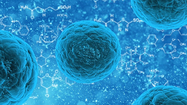
Like humans and living organisms, a body cell also goes through a life course of its own. It passes through several stages, from the time of its birth from a mother cell until the time it gives birth to two daughter cells. Our body cells have varying lengths of cell cycles but usually take around 24 hours to complete one course.
To know exactly a cell’s “age” during the course of its life cycle is one crucial aspect that interests scientists. But this has not been done in the past on living cells given that it’s only possible to know a cell’s age if it’s already dead. But a team of researchers from New York University (NYU) was able to do just that by using a state-of-the-art technique called fluorescence microscopy.
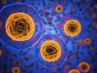
In eukaryotic cells (nucleus-containing cells), the cell cycle is composed of two phases. First is the interphase, the stage when the cell grows and makes a copy of its DNA in preparation for mitotic phase. The second is the mitotic phase or mitosis; a cell undergoes division to give birth to two identical daughter cells.
The cell cycle is indeed a cycle—it’s not just a linear timeline since at the end the daughter cells can start the same process all over again. These cells can either enter interphase immediately, or exit the cell cycle to step into a resting or “quiescent” phase. Daughter cells don’t actually, and as the term suggests, rest here. In fact, they continue to perform their designated jobs. For example, neurons conduct signals or liver cells store energy. The cells can be stuck in the resting phase permanently but, if signaled, they can enter a new cell cycle.
When a cell goes through its cell cycle, the size and shape of its nucleus undergoes big changes. Catching though the small and dynamic transitions can be tricky and difficult. To do that, NYU researchers used fluorescence microscopy to detect the flickers of the nuclear envelope, which last for just a few seconds (in fluorescence microscopy, the specimen are treated with a fluorescing material and then observed under a microscope).
The flickers decrease with the cell’s age – the older a cell , the weaker these flickers appear – and this observable quality is being utilized as a reliable indicator of the cell cycle stage. Alexandra Zidovska, the study’s senior author and a physics professor at NYU, explains, “this process can serve as an internal clock of the cell, telling you at what stage in the cell cycle the cell is.”
Why is it important to determine the cell’s stage in its cell cycle? Researchers hope that by learning to read a cell’s “internal clock”, we can gain a better understanding of the human biology, and improve our knowledge of both healthy and diseased cells.
“We know that structural and functional errors of the nuclear envelope lead to a large number of developmental and inherited disorders, such as cardiomyopathy, muscular dystrophy, and cancer. Illuminating the mechanics of nuclear shape fluctuations might contribute to efforts to understand the nuclear envelope in health and disease,” Zidovska added.
Details of the research can be viewed through the journal Proceedings of the National Academy of Sciences of the United States of America.
Disclaimer: This page contains affiliate links. If you choose to make a purchase after clicking a link, we may receive a commission at no additional cost to you. Thank you for your support!
Leave a Reply