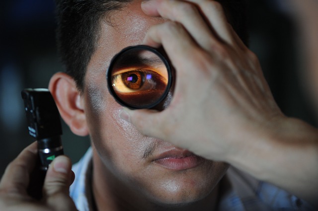
If you’ve ever gone through an eye exam before, you know how totally uncomfortable those eye drops are. They’re not that easy to put in; they sting; and they mess up your vision for a few more hours after you’re done with the exam. But, they’re an integral part of the process because without those drops, one’s pupils will instinctively close when exposed to a strong light source, which is exactly what’s needed to have your eyes properly checked.
With the latest camera being developed by a team of researchers from the University of Illinois and Harvard Medical School/Massachusetts Eye & Ear, those pupil-dilating eye drops may eventually become obsolete.
As it is, most retina cameras today make use of white light which causes the pupil to close, necessitating the use of eye drops. In contrast, this new retina camera works by combining harmless infrared light (which won’t make the pupil close because the iris doesn’t react to it) and white light. Infrared light will be used to focus light on the retina, and once focused, a brief flash of white light will be delivered as the image is being captured.
The team says that their prototype camera is made of simple parts that are readily available and only costs about $185.00 to make. It’s also quite small that it can be easily carried in one’s pocket. More importantly, it lets a doctor take pictures of the back of the eyes, without the need for eye drops. Afterwards, they can easily share those images with other doctors, and attach a copy to the patient’s medical record.
While there are already existing cameras that make use of the infrared/white light combination, they’re bulky and quite expensive. Which is what classifies this new camera a significant breakthrough.
According to Dr. Bailey Shen, an ophthalmology and visual sciences resident at the UIC College of Medicine and one of the authors of the research, the photos taken by their camera shows the retina, its blood supply, and the part of the optic nerve leading to the retina. Together, these images can be used to detect issues like diabetes, glaucoma and even elevated pressure around the brain.
Although the device is merely a prototype at this time, it proves that a camera that takes high quality pictures does not necessarily have to be expensive. Even better, it shows that it is definitely possible to do away with those eye drops.
As reported in UIC News, Dr. Shizuo Mukai, co-author of the study, retina surgeon at Massachusetts Eye and Ear, and a professor of ophthalmology at Harvard Medical School, said: “This is an open-source device that is cheap and easy to build. We expect that others who build our camera will add their own improvements and innovations.”
A description of the camera and how it can be assembled is available online through the Journal of Ophthalmology.
- Bulenox: Get 45% to 91% OFF ... Use Discount Code: UNO
- Risk Our Money Not Yours | Get 50% to 90% OFF ... Use Discount Code: MMBVBKSM
Disclaimer: This page contains affiliate links. If you choose to make a purchase after clicking a link, we may receive a commission at no additional cost to you. Thank you for your support!

Leave a Reply